欢迎访问《日用化学工业(中英文)》,今天是


日用化学工业(中英文) ›› 2026, Vol. 56 ›› Issue (4): 529-538.doi: 10.3969/j.issn.2097-2806.2026.04.013
王峥1,钱佳俊1,樊雨梅1,凌沛学1,2,*( )
)
收稿日期:2025-12-04
修回日期:2026-04-09
出版日期:2026-04-22
发布日期:2026-05-27
Zheng Wang1,Jiajun Qian1,Yumei Fan1,Peixue Ling1,2,*()
Received:2025-12-04
Revised:2026-04-09
Online:2026-04-22
Published:2026-05-27
Contact:
* E-mail: 摘要:
硫酸皮肤素(Dermatan sulfate, DS)是一种由N-乙酰半乳糖胺(GalNAc)与糖醛酸交替连接而成的糖胺聚糖,具有显著的结构异质性和高负电荷密度,能够与多种蛋白质发生特异性相互作用,广泛参与组织发育、损伤修复、炎症调控等关键生理与病理过程。皮肤中,DS通过调控胶原纤维组装、影响细胞信号传导和介导细胞黏附,在维持皮肤机械强度、促进伤口愈合和抗衰老等方面发挥关键作用。近年来,随着对DS生物学功能研究的深入,其在皮肤健康与疾病干预领域的应用潜力日益凸显。本文系统梳理了DS在皮肤组织工程、创面愈合、抗纤维化和抗老化等前沿领域的最新研究进展,剖析了DS构象灵活性(由艾杜糖醛酸介导)及其特定硫酸化模式如何调控皮肤生物功能的核心机制,以期为相关基础研究的转化与创新产品开发提供理论依据和新思路。
中图分类号:
王峥, 钱佳俊, 樊雨梅, 凌沛学. 硫酸皮肤素在皮肤组织中的生理功能与应用[J]. 日用化学工业(中英文), 2026, 56(4): 529-538.
Zheng Wang, Jiajun Qian, Yumei Fan, Peixue Ling. Physiological functions and applications of dermatan sulfate in skin tissue[J]. China Surfactant Detergent & Cosmetics, 2026, 56(4): 529-538.
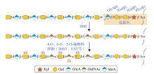
图1
DS生物合成途径示意图[10]"
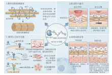
图2
DCN的皮肤生理功能"

表1
DS在皮肤医学领域的应用"
| 来源 | 结构特征 | 优势 | 劣势 | 主要应用场景 |
|---|---|---|---|---|
| 从陆地哺乳动物(猪、牛等)组织中提取 | 以单硫酸化单元为主,分子量相对较低,分布范围较窄 | 免疫原性弱、生产工艺成熟 | 硫酸化程度有限,靶点特异性较低 | 皮肤组织工程、抗衰老、抗炎、保湿产品 |
| 从海洋动物(鲨鱼、鳐鱼、三文鱼等)组织中提取 | 以双硫酸化或三硫酸化二糖单元为主,结构复杂,常与CS形成杂合链,分子量分布范围较宽 | 硫酸化模式复杂,具有多靶点效果;不存在宗教限制 | 提取纯化工艺复杂,成本较高 | 经皮医疗设备 |
| 人工重组DS(通过基因工程、酶催化合成或微生物表达系统生产) | 可设计特定硫酸化位点,分子量分布均一 | 纯度高、无动物源污染 | 研发成本较高 | 目前应用较为有限 |
| [1] |
Meyer K, Chaffee E. The mucopolysaccharides of skin[J]. Journal of Biological Chemistry, 1941, 138(2): 491-499.
doi: 10.1016/S0021-9258(18)51374-0 |
| [2] |
Silipo A, Zhang Z Q, Cañada F J, et al. Conformational analysis of a dermatan sulfate-derived tetrasaccharide by NMR, molecular modeling, and residual dipolar couplings[J]. Chembiochem, 2008, 9(2): 240-252.
doi: 10.1002/cbic.200700400 pmid: 18072186 |
| [3] | Wang W S, Shi L R, Qin Y, et al. Research and application of chondroitin sulfate/dermatan sulfate-degrading enzymes[J]. Frontiers in Cell and Developmental Biology, 2020. DOI: 10.3389/fcell.2020.560442. |
| [4] |
Habuchi O. Functions of chondroitin/dermatan sulfate containing GalNAc4, 6-disulfate[J]. Glycobiology, 2022, 32(8): 664-678.
doi: 10.1093/glycob/cwac030 pmid: 35552694 |
| [5] | Wisowski G, Paul-Samojedny M, Komosinska-Vassev K, et al. Structural variants of dermatan sulfate can affect the expression of proteins involved in breast cancer cell survival[J]. Cells, 2025, 14(20). DOI: 10.3390/cells14201581. |
| [6] |
Zhu J, Li Y, Shen W, et al. Relationships between transforming growth factor-β1, myostatin, and decorin: Implications for skeletal muscle fibrosis[J]. Journal of Biological Chemistry, 2007, 282(35): 25852-25863.
doi: 10.1074/jbc.M704146200 |
| [7] | Mahoutforoush A, Rasoulzadehzali M, Ashrafi E, et al. Dermatan sulfate-recognized UiO-66 metal-organic frameworks: An active and passive targeting nanomedicine for methotrexate delivery[J]. International Journal of Biological Macromolecules, 2025. DOI: 10.1016/j.ijbiomac.2025.148115. |
| [8] | Walimbe T, Panitch A. Proteoglycans in biomedicine: resurgence of an underexploited class of ECM molecules[J]. Frontiers in Pharmacology, 2020. DOI: 10.3389/fphar.2019.01661. |
| [9] | Yuan Q X, Shi X, Ma H Q, et al. Recent progress in marine chondroitin sulfate, dermatan sulfate, and chondroitin sulfate/dermatan sulfate hybrid chains as potential functional foods and therapeutic agents[J]. International Journal of Biological Macromolecules, 2024. DOI: 10.1016/j.ijbiomac.2024.129969. |
| [10] | Chen C C, Zhang X Y, Zhang W T, et al. Dermatan sulfate: structure, biosynthesis, and biological roles[J]. Biomolecules, 2025, 15(8). DOI: 10.3390/biom15081158. |
| [11] |
Port H, Andelic M, Nyström A, et al. Extracellular matrix alterations in inflammatory skin diseases: emerging biomarkers and clinical implications[J]. Clinical and Experimental Dermatology, 2025, 50(11): 2118-2132.
doi: 10.1093/ced/llaf308 pmid: 40632820 |
| [12] | Zhang B, Chi L I. Chondroitin sulfate/dermatan sulfate-protein interactions and their biological functions in human diseases: implications and analytical tools[J]. Frontiers in Cell and Developmental Biology, 2021. DOI: 10.3389/fcell.2021.693563. |
| [13] | Catarino C M, Kaiser K, Baltazar T, et al. Evaluation of native and non-native biomaterials for engineering human skin tissue[J]. Bioengineering & Translational Medicine, 2022, 7(3). DOI: 10.1002/btm2.10297. |
| [14] |
Mizumoto S. Defects in biosynthesis of glycosaminoglycans cause hereditary bone, skin, heart, immune, and neurological disorders[J]. Trends in Glycoscience and Glycotechnology, 2018, 30(174): 67-89.
doi: 10.4052/tigg.1812.2E |
| [15] | Mizumoto S, Yamada S, Sugahara K. Mutations in biosynthetic enzymes for the protein linker region of chondroitin/dermatan/heparan sulfate cause skeletal and skin dysplasias[J]. Biomed Research International, 2015. DOI: 10.1155/2015/861752. |
| [16] | Palhares L, London J A, Kozlowski A M, et al. Chemical modification of glycosaminoglycan polysaccharides[J]. Molecules, 2021, 26(17). DOI: 10.3390/molecules26175211. |
| [17] | Mizumoto S, Yamada S. Histories of dermatan sulfate epimerase and dermatan 4-o-sulfotransferase from discovery of their enzymes and genes to musculocontractural ehlers-danlos syndrome[J]. Genes, 2023, 14(2). DOI: 10.3390/genes14020509. |
| [18] | Sarbu M, Ica R, Sharon E, et al. Identification and structural characterization of novel chondroitin/dermatan sulfate hexassacharide domains in human decorin by ion mobility tandem mass spectrometry[J]. Molecules, 2022, 27(18). DOI: 10.3390/molecules27186026. |
| [19] | Mizumoto S, Yamada S. An overview of in vivo functions of chondroitin sulfate and dermatan sulfate revealed by their deficient mice[J]. Frontiers in Cell and Developmental Biology, 2021. DOI: 10.3389/fcell.2021.764781. |
| [20] |
Stojnic B, Galmés S, Serrano A, et al. Glycosaminoglycan dermatan sulfate supplementation decreases diet-induced obesity and metabolic dysfunction in mice[J]. Biofactors, 2024, 50(3): 493-508.
doi: 10.1002/biof.v50.3 |
| [21] |
Raman R, Sasisekharan V, Sasisekharan R. Structural insights into biological roles of protein-glycosaminoglycan interactions[J]. Chemistry & Biology, 2005, 12(3): 267-277.
doi: 10.1016/j.chembiol.2004.11.020 |
| [22] |
Nomura Y. Structural change in decorin with skin aging[J]. Connective Tissue Research, 2006, 47(5): 249-255.
pmid: 17118746 |
| [23] |
Taylor K R, Rudisill J A, Gallo R L. Structural and sequence motifs in dermatan sulfate for promoting fibroblast growth factor-2 (FGF-2) and FGF-7 activity[J]. Journal of Biological Chemistry, 2005, 280(7): 5300-5306.
doi: 10.1074/jbc.M410412200 pmid: 15563459 |
| [24] |
Kozma E M, Wisowski G, Kusz D, et al. The role of decorin and biglycan dermatan sulfate chain(s) in fibrosis-affected fascia[J]. Glycobiology, 2011, 21(10): 1301-1316.
doi: 10.1093/glycob/cwr065 pmid: 21543445 |
| [25] | Li Y, Liu Y, Xia W, et al. Age-dependent alterations of decorin glycosaminoglycans in human skin[J]. Scientific Reports, 2013. DOI: 10.1038/srep02422. |
| [26] | Baghy K, Szakadáti H, Kovalszky I. Decorin the antifibrotic proteoglycan and its progression in therapy[J]. American Journal of Physiology-Cell Physiology, 2025, 328(6): 1853-1865. |
| [27] |
Feugaing D D S, Götte M, Viola M. More than matrix: The multifaceted role of decorin in cancer[J]. European Journal of Cell Biology, 2013, 92(1): 1-11.
doi: 10.1016/j.ejcb.2012.08.004 pmid: 23058688 |
| [28] | Silver F H, Kelkar N, Deshmukh T. Molecular basis for mechanical properties of ecms: Proposed role of fibrillar collagen and proteoglycans in tissue biomechanics[J]. Biomolecules, 2021, 11(7). DOI: 10.3390/biom11071018. |
| [29] | Schmidt G, Hausser H, Kresse H. Interaction of the small proteoglycan decorin with fibronectin. Involvement of the sequence NKISK of the core protein[J]. Biochem J, 1991, 280(Pt 2): 411-414. |
| [30] |
Scott J. Proteoglycan-fibrillar collagen interactions[J]. Biochem J, 1988, 252(2): 313-323.
pmid: 3046606 |
| [31] | Mizumoto S, Yamada S. The specific role of dermatan sulfate as an instructive glycosaminoglycan in tissue development[J]. International Journal of Molecular Sciences, 2022, 23(13). DOI: 10.3390/ijms23137485. |
| [32] |
Cortes-Medina M, Bushman A R, Beshay P E, et al. Chondroitin sulfate, dermatan sulfate, and hyaluronic acid differentially modify the biophysical properties of collagen-based hydrogels[J]. Acta Biomaterialia, 2024, 174: 116-126.
doi: 10.1016/j.actbio.2023.12.018 pmid: 38101556 |
| [33] |
Eremenko E, Ding J, Kwan P, et al. The biology of extracellular matrix proteins in hypertrophic scarring[J]. Advances in Wound Care, 2022, 11(5): 234-254.
doi: 10.1089/wound.2020.1257 |
| [34] | Hashimoto A, Hirose T, Hashimoto K, et al. Collagen network formation in in vitro models of musculocontractural ehlers-danlos syndrome[J]. Genes, 2023, 14(2). DOI: 10.3390/genes14020308. |
| [35] | Tota C G, Leone A, Khan A, et al. Ccant1 affects cartilage proteoglycan properties: aggrecan and decorin characterization in a mouse model of desbuquois dysplasia type 1[J]. Biomolecules, 2024, 14(9). DOI: 10.3390/biom14091064. |
| [36] | Huang J W, Heng S J, Zhang W L, et al. Dermal extracellular matrix molecules in skin development, homeostasis, wound regeneration and diseases[J]. Seminars in Cell & Developmental Biology, 2022, 128: 137-144. |
| [37] |
Kuwaba K, Kobayashi M, Nomura Y, et al. Size control of decorin dermatan sulfate during remodeling of collagen fibrils in healing skin[J]. Journal of Dermatological Science, 2002, 29(3): 185-194.
pmid: 12234708 |
| [38] |
Iwasaki S, Hosaka Y, Iwasaki T, et al. The modulation of collagen fibril assembly and its structure by decorin: An electron microscopic study[J]. Archives of Histology and Cytology, 2008, 71(1): 37-44.
pmid: 18622092 |
| [39] |
Rüehland C, Schönherr E, Robenek H, et al. The glycosaminoglycan chain of decorin plays an important role in collagen fibril formation at the early stages of fibrillogenesis[J]. Febs Journal, 2007, 274(16): 4246-4255.
doi: 10.1111/j.1742-4658.2007.05951.x pmid: 17651433 |
| [40] |
Concha M, Vidal A, Giacaman A, et al. Aerogels made of chitosan and chondroitin sulfate at high degree of neutralization: Biological properties toward wound healing[J]. Journal of Biomedical Materials Research Part B-Applied Biomaterials, 2018, 106(6): 2464-2471.
doi: 10.1002/jbm.b.34038 pmid: 29424958 |
| [41] | Mizumoto S, Kosho T, Yamada S, et al. Pathophysiological significance of dermatan sulfate proteoglycans revealed by human genetic disorders[J]. Pharmaceuticals, 2017, 10(2). DOI: 10.3390/ph10020034. |
| [42] |
Zhang Z, Garron T, Li X J, et al. Recombinant human decorin inhibits TGF-β1 induced contraction of collagen lattice by keloid fibroblasts[J]. Wounds-a Compendium of Clinical Research and Practice, 2009, 21(2): 47-56.
pmid: 25903025 |
| [43] | Takaya K, Aramaki-Hattori N, Sakai S, et al. Decorin inhibits dermal mesenchymal cell migration and induces scar formation[J]. Plastic and Reconstructive Surgery-Global Open, 2022, 10(4). DOI: 10.1097/gox.0000000000004245. |
| [44] |
Radek K, Taylor K, Gallo R. FGF-10 and specific structural elements of dermatan sulfate size and sulfation promote maximal keratinocyte migration and cellular proliferation[J]. Wound Repair and Regeneration, 2009, 17(1): 118-126.
doi: 10.1111/j.1524-475X.2008.00449.x pmid: 19152659 |
| [45] |
Lochner K, Gaemlich A, Suedel K M, et al. Expression of decorin and collagens I and III in different layers of human skin in vivo: A laser capture microdissection study[J]. Biogerontology, 2007, 8(3): 269-282.
pmid: 17146610 |
| [46] | Yan Y, Quan H H, Guo C F, et al. Alterations of matrisome gene expression in naturally aged and photoaged human skin in vivo[J]. Biomolecules, 2024, 14(8). DOI: 10.3390/biom14080900. |
| [47] | Pang X X, Dong N, Zheng Z. Small leucine-rich proteoglycans in skin wound healing[J]. Frontiers in Pharmacology, 2020. DOI: 10.3389/fphar.2019.01649. |
| [48] | Wadstein J, Alvarez I S, López L B. Managing skin ageing as a modifiable disorder-the clinical application of nourella® dual approach comprising a nano-encapsulated retinoid, retiliex-a® and a skin proteoglycan replacement therapy, vercilex®[J]. Cosmetics. 2022, 9(2): DOI: 10.3390/cosmetics9020031. |
| [49] |
Lujan T J, Underwood C J, Jacobs N T, et al. Contribution of glycosaminoglycans to viscoelastic tensile behavior of human ligament[J]. Journal of Applied Physiology, 2009, 106(2): 423-431.
doi: 10.1152/japplphysiol.90748.2008 pmid: 19074575 |
| [50] |
Población C A, Michelacci Y M. Structural differences of dermatan sulfates from different origins[J]. Carbohydrate Research, 1986, 147(1): 87-100.
pmid: 3698057 |
| [51] |
Schnabelrauch M, Scharnweber D, Schiller J. Sulfated glycosaminoglycans as promising artificial extracellular matrix components to improve the regeneration of tissues[J]. Current Medicinal Chemistry, 2013, 20(20): 2501-2523.
pmid: 23521682 |
| [52] | Li K Q, Li R F, Liu Y J, et al. Diversity, mechanism and structure-activity relationships of marine anticoagulant-active polysaccharides: A review[J]. International Journal of Biological Macromolecules, 2025. DOI: 10.1016/j.ijbiomac.2025.141742. |
| [53] |
Ruozi B, Parma B, Croce M A, et al. Collagen-based modified membranes for tissue engineering: Influence of type and molecular weight of GAGs on cell proliferation[J]. International Journal of Pharmaceutics, 2009, 378(1-2): 108-115.
doi: 10.1016/j.ijpharm.2009.05.049 pmid: 19501149 |
| [54] | Chocarro-Wrona C, Pleguezuelos-Beltrán P, de Andrés J L, et al. A bioactive three-layered skin substitute based on ecm components effectively promotes skin wound healing and regeneration[J]. Materials Today Bio., 2025. DOI: 10.1016/j.mtbio.2025.101592. |
| [55] | Chocarro-Wrona C, de Andrés J L, Rioboó-Legaspi P, et al. Design and evaluation of a bilayered dermal/hypodermal 3D model using a biomimetic hydrogel formulation[J]. Biomedicine & Pharmacotherapy, 2024. DOI: 10.1016/j.biopha.2024.117051. |
| [56] |
DeAngelis P L, Liu J, Linhardt R J. Chemoenzymatic synthesis of glycosaminoglycans: Re-creating, re-modeling and re-designing nature’s longest or most complex carbohydrate chains[J]. Glycobiology, 2013, 23(7): 764-777.
doi: 10.1093/glycob/cwt016 pmid: 23481097 |
| [57] |
Nillesen S, Lammers G, Wismans R, et al. Design and in vivo evaluation of a molecularly defined acellular skin construct: Reduction of early contraction and increase in early blood vessel formation[J]. Acta Biomaterialia, 2011, 7(3): 1063-1071.
doi: 10.1016/j.actbio.2010.10.011 pmid: 20965285 |
| [58] | Pleguezuelos-Beltrán P, Nieto-García D, Chocarro-Wrona C, et al. A novel sprayable fibrinogen/glycosaminoglycans/collagen-based bioink for skin wound healing applied by a handheld dual-head airbrush[J]. Advanced Healthcare Materials, 2025. DOI: 10.1002/adhm.202500702. |
| [59] |
He S Q, Walimbe T, Chen H Y, et al. Bioactive extracellular matrix scaffolds engineered with proangiogenic proteoglycan mimetics and loaded with endothelial progenitor cells promote neovascularization and diabetic wound healing[J]. Bioactive Materials, 2022, 10: 460-473.
doi: 10.1016/j.bioactmat.2021.08.017 pmid: 34901560 |
| [60] |
Sato E, Zhang L J, Dorschner R, et al. Activation of parathyroid hormone 2 receptor induces decorin expression and promotes wound repair[J]. Journal of Investigative Dermatology, 2017, 137(8): 1774-1783.
doi: S0022-202X(17)31424-0 pmid: 28454729 |
| [61] | Olczyk P, Komosinska-Vassev K, Winsz-Szczotka K, et al. Propolis induces chondroitin/dermatan sulphate and hyaluronic acid accumulation in the skin of burned wound[J]. Evidence-Based Complementary and Alternative Medicine, 2013. DOI: 10.1155/2013/290675. |
| [62] |
Zhang Z, Li X J, Liu Y, et al. Recombinant human decorin inhibits cell proliferation and downregulates TGF-β1 production in hypertrophic scar fibroblasts[J]. Burns, 2007, 33(5): 634-641.
pmid: 17374457 |
| [63] |
Pemmari T, Ivanova L, May U, et al. Exposed cendr domain in homing peptide yields skin-targeted therapeutic in epidermolysis bullosa[J]. Molecular Therapy, 2020, 28(8): 1833-1845.
doi: S1525-0016(20)30251-3 pmid: 32497513 |
| [64] |
Veettil S P, Gopinath A, Madhan B, et al. A cyclodextrin-based macrocyclic oligosaccharide cavitand with a dual functionality limits the collagen fibrillogenesis: A possible carbohydrate-based therapeutic molecule for fibrotic diseases[J]. International Journal of Biological Macromolecules, 2022, 207: 222-231.
doi: 10.1016/j.ijbiomac.2022.03.005 pmid: 35259432 |
| [65] | Galvez-Martin P, Martinez-Puig D, Soto-Fernández C, et al. In vitroevaluation of anti-aging and regenerative properties of dermatan sulfate for skin care[J]. The FASEB Journal, 2022, 36(S1). DOI: 10.1096/fasebj.2022.36.S1.L7681. |
| [66] |
Reinboth B, Hanssen E, Cleary E G, et al. Molecular interactions of biglycan and decorin with elastic fiber components-Biglycan forms a ternary complex with tropoelastin and microfibril-associated glycoprotein 1[J]. Journal of Biological Chemistry, 2002, 277(6): 3950-3957.
doi: 10.1074/jbc.M109540200 pmid: 11723132 |
| [67] | Segarra S, Bosnjak I, Mioc I, et al. Yeast-Derived nucleotides enhance fibroblast migration and proliferation and provide clinical benefits in atopic dermatitis[J]. International Journal of Molecular Sciences, 2024, 25(5). DOI: 10.3390/ijms25052890. |
| [68] | Vassal-Stermann E, Duranton A, Black A, et al. A new c-xyloside induces modifications of gag expression, structure and functional properties[J]. Plos One, 2012, 7(10). DOI: 10.1371/journal.pone. 0047933. |
| [69] | Betts R J, 苏士纹, 孔佩慧, 等. 局部外用C-木糖苷促进皮肤年轻化的研究进展[J]. 临床皮肤科杂志, 2021, 50(7): 444-448. |
| [70] |
Peramo A, Marcelo C L, Goldstein S A, et al. Improved preservation of the tissue surrounding percutaneous devices by hyaluronic acid and dermatan sulfate in a human skin explant model[J]. Annals of Biomedical Engineering, 2010, 38(3): 1098-1110.
doi: 10.1007/s10439-009-9872-1 pmid: 20020211 |
| [71] | Belvedere R, Bizzarro V, Parente L, et al. The pharmaceutical device prisma® skin promotes in vitro angiogenesis through endothelial to mesenchymal transition during skin wound healing[J]. International Journal of Molecular Sciences, 2017, 18(8). DOI: 10.3390/ijms18081614. |
| [72] |
Dhahri M, Abed A, Lajimi R H, et al. Grafting of dermatan sulfate on polyethylene terephtalate to enhance biointegration[J]. Journal of Biomedical Materials Research Part A, 2011, 98(1): 114-121.
doi: 10.1002/jbm.a.33077 pmid: 21548062 |
| [73] | 陈晨, 王冠凤, 石艳丽, 等. 一种含依克多因的组合物及其在激光美容中的应用: CN111714396A[P]. 2020-09-29. |
| [74] | Vega V L, Colvan L, Fleck T. Topical low molecular weight sulfated glycosaminoglycan analogs improve skin tightening and deep hydration in subjects with moderate to severe facial photodamage[J]. Journal of the American Academy of Dermatology, 2019, 81(4): 139. |
| [75] | Zhang L Y, Dong Z R, Liu W J, et al. Novel pharmaceutical strategies for enhancing skin penetration of biomacromolecules[J]. Pharmaceuticals, 2022, 15(7). DOI:10.3390/ph15070877. |
| [76] |
Paderi J E, Panitch A. Design of a synthetic collagen-binding peptidoglycan that modulates collagen fibrillogenesis[J]. Biomacromolecules, 2008, 9(9): 2562-2566.
doi: 10.1021/bm8006852 pmid: 18680341 |
| [77] |
Lyon M, Deakin J A, Rahmoune H, et al. Hepatocyte growth factor scatter factor binds with high affinity to dermatan sulfate[J]. Journal of Biological Chemistry, 1998, 273(1): 271-278.
doi: 10.1074/jbc.273.1.271 pmid: 9417075 |
| [1] | 高欣雨, 孙娅楠, 李俐霜, 金玉翠, 马淑骅, 王毅. 东阿阿胶对小鼠皮肤屏障损伤的修复作用研究[J]. 日用化学工业(中英文), 2026, 56(2): 236-244. |
| [2] | 王丹羽, 王学硕, 骆海朋, 李景云, 崔生辉, 路勇. 人面部皮肤可培养细菌的物种多样性及化妆品防腐剂的影响研究[J]. 日用化学工业(中英文), 2026, 56(2): 208-216. |
| [3] | 李佳明, 邱晓锋, 郭惠珊, 罗建锟, 甘礼社, 徐学涛. 松茸蛋白的制备、结构表征及其保湿功效研究[J]. 日用化学工业(中英文), 2026, 56(1): 98-106. |
| [4] | 申亚利, 刘培林, 刘琳立, 范丽敏, 李焕英. 黑豆多肽对UVB诱导的人皮肤成纤维细胞活性和胶原分泌的影响[J]. 日用化学工业(中英文), 2025, 55(9): 1179-1186. |
| [5] | 贾东晗, 伍琰妍, 龚贺吉, 刘哲伟, 彭常梅, 张婉萍. 不同活性成分对唇部修护能力的评价[J]. 日用化学工业(中英文), 2025, 55(8): 1024-1034. |
| [6] | 刘敏, 叶磊, 蒋玲玲, 郑久炎. 神经酰胺脂质体对光损伤的修护及机械损伤的修复功效研究[J]. 日用化学工业(中英文), 2025, 55(7): 902-908. |
| [7] | 刘婷, 刘师卜, 张励, 朵慧, 路勇, 张露勇. 皮肤致敏体外方法:动力学直接多肽反应试验方法(kDPRA)的转化与验证[J]. 日用化学工业(中英文), 2025, 55(6): 723-730. |
| [8] | 吴凡, 张嘉琪, 秦毅, 王俊, 吴宗翰, 盘瑶. 化妆品活性成分通过细胞自噬发挥护肤功效的研究进展[J]. 日用化学工业(中英文), 2025, 55(6): 803-810. |
| [9] | 帖航, 李琦, 朱翠翠, 袁旻嘉, 徐亮. 磷脂酰胆碱包裹的白藜芦醇增强皮肤抗氧化和抗皱能力研究[J]. 日用化学工业(中英文), 2025, 55(6): 756-766. |
| [10] | 厉浩东, 吴兴怡, 张影, 王玮恋, 王伟, 李丽. 天麻素对UVB损伤皮肤的修复作用[J]. 日用化学工业(中英文), 2025, 55(6): 779-784. |
| [11] | 谢诗雅, 刘明巍, 肖雷, 胡绍霖. 蒲公英多酚的抑菌和抗炎活性研究[J]. 日用化学工业(中英文), 2025, 55(5): 633-639. |
| [12] | 苗开元, 欧阳泽垚, 赵新颖, 杨晓婧, 陈欣妍, 石召华. 氧化白藜芦醇自微乳的制备、表征及美白活性的研究[J]. 日用化学工业(中英文), 2025, 55(4): 453-463. |
| [13] | 杨武成, 谢宇, 魏剑, 范瑞芳, 谭建华, 席绍峰. 共聚焦拉曼光谱在皮肤屏障功能评估和经皮吸收研究中的应用[J]. 日用化学工业(中英文), 2025, 55(4): 508-515. |
| [14] | 李以洪, 许梦然, 盘瑶, 吴金昊, 刘琦, 常思思, 赵华. 基于KNN算法建立晒后皮肤状态评估模型[J]. 日用化学工业(中英文), 2025, 55(3): 349-357. |
| [15] | 赵化冰, 李颖甜, 王熙函, 黄正梅, 路福平. 皮肤微生态与微生态护肤品[J]. 日用化学工业(中英文), 2025, 55(3): 390-398. |
|
||
