欢迎访问《日用化学工业(中英文)》,今天是

日用化学工业 ›› 2020, Vol. 50 ›› Issue (8): 560-565.doi: 10.3969/j.issn.1001-1803.2020.08.009

• 开发与应用 • 上一篇    下一篇

柚皮苷对紫外线诱导小鼠皮肤屏障损伤的保护作用及机制研究

车飙1(),王静3,邓明高3,韩萍2,陈惠雄2,杜志云3()   

  1. 1.无限极(中国)有限公司,广东 江门 529156
    2.佛山市康伲爱伦生物技术有限公司,广东 佛山 528200
    3.广东工业大学 生物医药学院,广东 广州 510006
  • 收稿日期:2020-03-08 修回日期:2020-07-27 出版日期:2020-08-22 发布日期:2020-08-25
  • 通讯作者: 杜志云
  • 作者简介:车 飙(1968-),男,工程师,硕士,电话:13808821878,E-mail:bill.che@infinitus-int.com

Protective effect and mechanism of naringin on UV-induced skin barrier damage in mice

CHE Biao1(),WANG Jing3,DENG Ming-gao3,HAN Ping2,CHEN Hui-xiong2,DU Zhi-yun3()   

  1. 1. Infinitus (China) Co., Ltd, Jiangmen, Guangdong 529156, China
    2. Allan Conney Biotechnology Company Ltd, Foshan, Guangdong 528200, China
    3. School of Biomedical and Pharmaceutical Sciences, Guangdong University of Technology, Guangzhou, Guangdong 510006, China
  • Received:2020-03-08 Revised:2020-07-27 Online:2020-08-22 Published:2020-08-25
  • Contact: Zhi-yun DU

摘要:

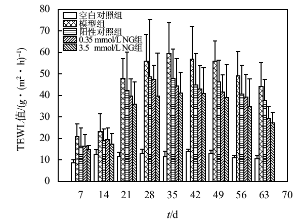
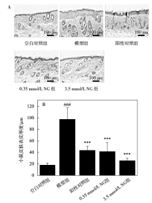
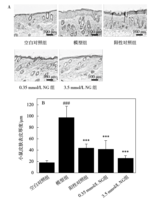
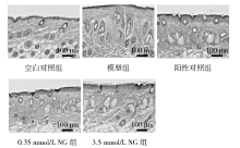
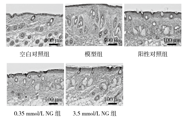
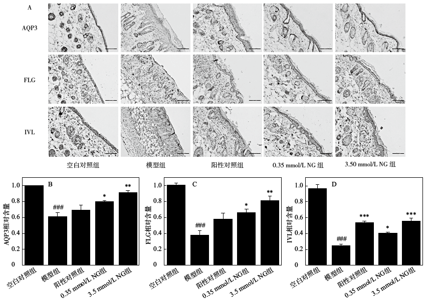
研究了柚皮苷对紫外线诱导昆明小鼠皮肤屏障损伤的保护作用。将25只SPF级昆明小鼠随机分为5组:空白对照组,模型组(UV照射组),阳性对照组(28.39 mmol/L维生素C),低、高剂量柚皮苷涂抹组(0.35和3.5 mmol/L柚皮苷)。除空白对照组,其他4组小鼠模拟日光紫外线(UVA + UVB)照射,建立小鼠光损伤皮肤模型,每次UV照射前,提前2 h分别涂抹相同剂量(100 μL/只)的药物。通过测试小鼠皮肤经表皮失水(TEWL)、H&E染色、胶原纤维染色(Masson染色),以及小鼠皮肤组织中的超氧化物歧化酶(SOD)、过氧化氢酶(CAT)的活力,丙二醛(MDA)的含量,活性氧(ROS)清除能力来探究柚皮苷对皮肤光损伤的作用途径;免疫组化染色(IHC染色)检测丝聚合蛋白(FLG)、内披蛋白(IVL)、水通道蛋白3 (AQP3)以评价柚皮苷对紫外线损伤皮肤的屏障相关功能。与模型组相比,柚皮苷涂抹组小鼠皮肤抑制皮肤表皮厚度增加,给药组胶原量增加;低、高剂量柚皮苷涂抹组可以显著减少光损伤小鼠皮肤中水分的流失(p<0.05);柚皮苷显著提高小鼠皮肤组织中SOD、CAT的活力(p<0.05),显著降低MDA的含量(p<0.05)及清除ROS的含量;IHC染色研究表明柚皮苷涂抹组皮肤中FLG、IVL、AQP3蛋白表达量增加(p<0.05)。

关键词: 柚皮苷, UV损伤, 皮肤屏障, 抗氧化

Abstract:

The protective effect of naringin on UV-induced skin barrier damage in KM mice was studied. Twenty-five SPF KM mice were randomly divided into 5 groups: blank control group, UV model group (UV irradiation group), positive control group (28.39 mmol/L vitamin C), low and high-dose naringin smear groups (0.35, 3.5 mmol/L naringin). Except for the blank control group, the other four groups of mice were simulated with sunlight ultraviolet (UVA+UVB) radiation to establish a mouse photo-damaged skin model. Before each UV irradiation, the same amount (100 μL/mouse) of naringin with the indicated dose was applied 2 h in advance. The effects of naringin on skin photodamage were investigated by testing the skin TEWL, H&E staining, Masson staining, the activities of superoxide dismutase (SOD) and catalase (CAT), malondialdehyde (MDA) content, and the scavenging ability of reactive oxygen species (ROS) in mice skin tissues. Moreover, immunohistochemical staining (IHC staining) of filaggrin (FLG), involucrin (IVL) and aquaporin 3 (AQP3) was used to evaluate the skin barrier-related functions of naringin against UV-damaged skin. Compared with the model group, the skin of the naringin administration group inhibited the increase in skin epidermal thickness, and the amount of collagen in the administration group increased; the low and high-dose naringin smear group could significantly reduce the loss of water in the skin of mice with UV damage (p<0.05). Naringin significantly increased the activity of SOD and CAT (p<0.05), significantly reduced the content of MDA (p<0.05), and eliminated the content of ROS in the skin of mice. IHC staining study showed that the protein expression level of FLG, IVL and AQP3 increased in the naringin-administered group (p<0.05).

Key words: naringin, UV damage, skin barrier, antioxidant

中图分类号: 

  • TQ658